What follows are two discrete photographs, each offering six individual arthroscopic images, organized in two equal columns of three, of Joseph P. Fisher’s right hip during labral repair surgery on May 31, 2023.
Series 1
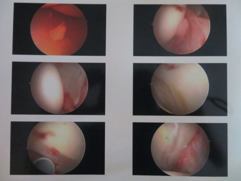
Image Description: a collection of six arthroscopic images of Joseph P. Fisher’s right hip taken during his labral repair surgery. Of particular note is the first image in the series located at the top of the left column. This image shows a small fragment of loose cartilage that was floating in the joint upon first inspection. In addition, the image at the bottom of the left column shows what is likely a cartilage defect on the cam portion of the femoral head. The defect is light red against the white articular cartilage. It is possible that the loose fragment in the joint broke loose from this part of the articular cartilage.
Series 2
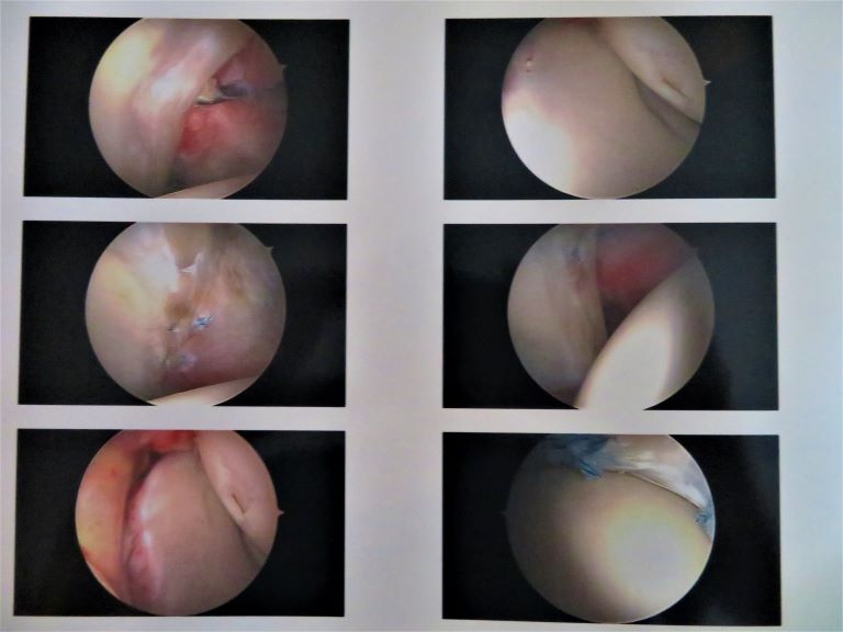
Image Description: six additional arthroscopic images of Joseph P. Fisher’s right hip taken during his labral repair surgery. The first and second images in the left column show the early stages of the labral suturing process. In each of these images pieces of thread in faint blue and white appear pulled between white cartilage. The bottom image in the right column, the sixth image in the series, shows a close-up of the blue and white thread bulled tight against smooth white articular cartilage. This image represents the completion of the repair procedure.